| ABAT |
HPA041690 |
Soma in neurons. |
| ACADL |
HPA011990 |
Soma and synapse in neurons. |
| ACO2 |
HPA001097 |
Soma and synapse in neurons. |
| ADCYAP1 |
HPA065887 |
Axon and synapse in neurons. |
| ADORA2A |
HPA065566 |
Soma, nucleus, dendrite and synapse in neurons. |
| AGRP |
HPA041017 |
Soma, dendrite, axon and synapse in neurons. |
| ALDH1A1 |
HPA002123 |
Endfeet in astrocytes. |
| ALDH1L1 |
HPA036900 |
Astrocytes. |
| ALDOC |
HPA067442 |
Soma in astrocytes. |
| AMPD2 |
HPA045760 |
Soma and synapse in neurons.
Circumventricular organs of ependymal cells. |
| AP000275.65 |
HPA028849 |
Soma and dendrite in neurons. |
| AQP4 |
HPA014784 |
Endfeet in astrocytes. |
| ARC |
CAB079013 |
|
| ARFGEF1 |
HPA023822 |
Soma and dendrite in neurons.
Ventricle wall and circumventricular organs of ependymal cells. |
| ARHGAP1 |
HPA004689 |
Soma and synapse in neurons. |
| AVP |
HPA071892 |
Soma and axon in neurons. |
| B3GNT6 |
HPA012158 |
Soma, dendrite and axon in neurons. |
| BBOX1 |
HPA027823 |
Soma in neurons. |
| BCAN |
HPA007865 |
Glia.
Soma and axon in neurons. |
| BCAP31 |
HPA003906 |
Synapse in neurons. |
| BCAR1 |
HPA042282 |
Soma and dendrite in neurons. |
| BCL11B |
HPA049117 |
Nucleus in neurons. |
| BCL2L2-PABPN1 |
HPA000637 |
Soma and synapse in neurons. |
| BIRC3 |
HPA002317 |
Soma, nucleus and dendrite in neurons.
Choroid plexus, ventricle wall and circumventricular organs of ependymal cells. |
| BMPER |
HPA018083 |
Soma, nucleus and synapse in neurons. |
| C12orf10 |
HPA038626 |
Soma and endfeet in astrocytes. |
| C17orf67 |
HPA043479 |
Dendrite and synapse in neurons. |
| C17orf75 |
HPA004061 |
Soma and dendrite in neurons. |
| C21orf59 |
HPA028849 |
Soma and dendrite in neurons. |
| CACNA1C |
CAB079031 |
Soma and synapse in neurons. |
| CACNA1E |
CAB079032 |
Synapse in neurons. |
| CALB2 |
HPA007305 |
Soma, dendrite and axon in neurons. |
| CALCA |
HPA059886 |
Soma and synapse in neurons. |
| CALCB |
HPA059886 |
Soma and synapse in neurons. |
| CAMK2B |
HPA026307 |
Soma and dendrite in neurons. |
| CAPN2 |
HPA024470 |
Soma and synapse in neurons.
Endothelia. |
| CASKIN1 |
CAB079050 |
Synapse in neurons. |
| CBLN3 |
HPA041266 |
Synapse in neurons. |
| CBLN4 |
HPA074497 |
Synapse in neurons. |
| CCDC22 |
HPA000229 |
Axon and synapse in neurons. |
| CCK |
HPA045039 |
Synapse in neurons. |
| CDC42EP4 |
HPA024797 |
Nucleus and dendrite in neurons. |
| CHGB |
HPA012602 |
Synapse in neurons. |
| CHODL |
HPA017282 |
Nucleus in neurons. |
| CIB2 |
HPA036697 |
Glia.
Soma, nucleus and dendrite in neurons. |
| CKS1B |
HPA003424 |
Soma in microglia.
Synapse in neurons.
Endothelia. |
| CKS2 |
HPA003424 |
Soma in microglia.
Synapse in neurons.
Endothelia. |
| CNGA3 |
CAB079025 |
Soma in neurons. |
| CNP |
HPA023338 |
Soma and myelin sheet in oligodendrocytes. |
| CPLX3 |
CAB079003 |
Axon and synapse in neurons. |
| CRABP1 |
HPA017203 |
Soma and synapse in neurons. |
| CRH |
HPA046846 |
Neurons. |
| CSK |
HPA028425 |
Soma and synapse in neurons. |
| CTBP2 |
CAB079018 |
Soma, nucleus and synapse in neurons.
Endothelia.
Choroid plexus, ventricle wall and circumventricular organs of ependymal cells. |
| CYGB |
HPA017757 |
Soma and dendrite in neurons. |
| DCX |
HPA036121 |
Soma and nucleus in neurons.
Ventricle wall of ependymal cells. |
| DDX3X |
HPA001648 |
Soma in neurons. |
| DDX3Y |
HPA001648 |
Soma in neurons. |
| DENND3 |
HPA041668 |
Soma, axon and synapse in neurons. |
| DGKB |
HPA020321 |
Nucleus, dendrite and synapse in neurons. |
| DNAJC30 |
HPA017318 |
Myelin sheet in oligodendrocytes. |
| DPP6 |
HPA050509 |
Soma and dendrite in neurons. |
| DRD1 |
HPA013393 |
Soma and synapse in neurons. |
| DRD2 |
HPA015139 |
Soma, dendrite and synapse in neurons. |
| DTX4 |
HPA059294 |
Soma in neurons.
Choroid plexus of ependymal cells. |
| ECEL1 |
HPA069756 |
Soma in neurons. |
| ECH1 |
HPA005835 |
Soma in astrocytes.
Soma and dendrite in neurons.
Endothelia.
Choroid plexus and circumventricular organs of ependymal cells. |
| EDN3 |
HPA036618 |
Glia.
Axon and synapse in neurons.
Ependymal cells. |
| EFCAB12 |
HPA037694 |
Neurons. |
| EFHC2 |
HPA034492 |
Soma, dendrite, axon and synapse in neurons. |
| EIF1AX |
HPA002561 |
Soma, nucleus and dendrite in neurons.
Choroid plexus, ventricle wall and circumventricular organs of ependymal cells. |
| EIF1AY |
HPA002561 |
Soma, nucleus and dendrite in neurons.
Choroid plexus, ventricle wall and circumventricular organs of ependymal cells. |
| EMX2 |
HPA003497 |
Dendrite, axon and synapse in neurons. |
| ENO2 |
CAB079023 |
Soma and synapse in neurons. |
| FAM213B |
HPA006403 |
Soma, dendrite and synapse in neurons. |
| FEV |
HPA067679 |
Soma, dendrite and axon in neurons.
Endothelia. |
| FGF3 |
HPA012692 |
Dendrite and synapse in neurons. |
| FH |
HPA025770 |
Soma and nucleus in neurons. |
| FKBP7 |
HPA008707 |
Soma, dendrite and synapse in neurons. |
| FLNA |
HPA001016 |
Synapse in neurons. |
| FOXO1 |
HPA001252 |
Soma in neurons. |
| FRMD4B |
HPA009705 |
Soma and dendrite in neurons. |
| FRMD6 |
HPA001297 |
Soma and nucleus in neurons.
Choroid plexus and ventricle wall of ependymal cells. |
| GABBR2 |
CAB079065 |
Soma, dendrite, axon and synapse in neurons. |
| GABRA2 |
CAB079061 |
Soma and synapse in neurons. |
| GABRA3 |
HPA000839 |
Soma, dendrite and synapse in neurons. |
| GABRA5 |
CAB079022 |
Soma and synapse in neurons. |
| GABRG2 |
CAB079060 |
Soma and synapse in neurons. |
| GAD1 |
CAB078176 |
Soma, dendrite, axon and synapse in neurons. |
| GAL |
HPA049864 |
Soma, dendrite and axon in neurons. |
| GFAP |
HPA056030 |
Soma in astrocytes. |
| GJA1 |
HPA069245 |
Astrocytes.
Synapse in neurons. |
| GKAP1 |
HPA035117 |
Soma and dendrite in neurons. |
| GLRA1 |
CAB079042 |
Axon and synapse in neurons. |
| GMFB |
HPA002954 |
Soma and dendrite in neurons. |
| GMFG |
HPA002954 |
Soma and dendrite in neurons. |
| GNG2 |
HPA003534 |
Synapse in neurons. |
| GOLGA5 |
HPA000992 |
Soma in neurons. |
| GPR17 |
HPA029766 |
Soma and nucleus in oligodendrocytes.
Soma in neurons. |
| GPR88 |
HPA007488 |
Axon and synapse in neurons. |
| GRM2 |
HPA027868 |
Axon and synapse in neurons. |
| GUCA1A |
HPA005561 |
Soma and synapse in neurons. |
| HAPLN1 |
HPA025238 |
Soma in neurons. |
| HOMER2 |
CAB079014 |
Soma and synapse in neurons. |
| HOMER3 |
CAB079015 |
Soma and synapse in neurons. |
| HPF1 |
HPA043467 |
Synapse in neurons. |
| HSPA2 |
HPA000798 |
Soma and nucleus in neurons. |
| IER5 |
HPA029894 |
Synapse in neurons. |
| IGDCC4 |
HPA008510 |
Synapse in neurons. |
| INA |
HPA008057 |
Soma, dendrite and axon in neurons. |
| IPO7 |
HPA019002 |
Soma in neurons. |
| ITIH3 |
HPA017373 |
Soma and nucleus in neurons. |
| ITPKA |
HPA040454 |
Soma and endfeet in astrocytes. |
| KCNIP4 |
HPA022862 |
Synapse in neurons. |
| KCTD12 |
HPA077376 |
Soma in neurons. |
| KIF5A |
HPA004469 |
Soma in neurons. |
| KIT |
HPA073252 |
Synapse in neurons. |
| KMT2E |
HPA022812 |
Neurons. |
| LEP |
HPA030721 |
Synapse in neurons. |
| LHX2 |
HPA000838 |
Soma in neurons. |
| LIAS |
HPA018842 |
Glia.
Soma, nucleus and dendrite in neurons.
Choroid plexus, ventricle wall and circumventricular organs of ependymal cells. |
| LPCAT2 |
HPA008433 |
Microglia. |
| LPO |
HPA028688 |
Soma and nucleus in oligodendrocytes. |
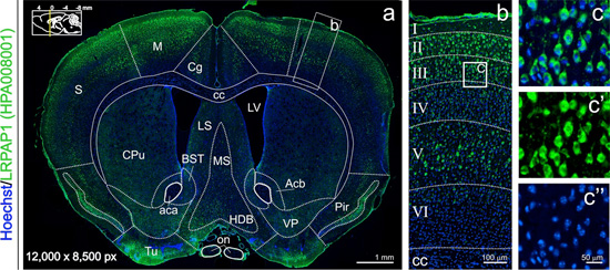
| LRPAP1 |
HPA008001 |
Soma and dendrite in neurons.
Choroid plexus and circumventricular organs of ependymal cells. |
| MAP2 |
HPA012828 |
Soma and dendrite in neurons. |
| MAPK6 |
HPA030262 |
Soma in neurons. |
| MARS |
HPA004125 |
Soma, dendrite and axon in neurons. |
| MBP |
HPA049222 |
Soma and myelin sheet in oligodendrocytes. |
| MCC |
HPA037390 |
Soma in astrocytes.
Soma in neurons. |
| MIA |
HPA031950 |
Soma in oligodendrocytes.
Soma in neurons. |
| MPP4 |
CAB079019 |
Synapse in neurons. |
| MROH1 |
HPA044465 |
Axon in neurons. |
| MTX2 |
HPA031550 |
Astrocytes. |
| MX1 |
HPA030918 |
Soma in astrocytes.
Soma in neurons. |
| NAGLU |
HPA038815 |
Soma, dendrite and synapse in neurons.
Circumventricular organs of ependymal cells. |
| NCDN |
HPA023676 |
Soma and dendrite in neurons. |
| NCS1 |
HPA019713 |
Dendrite, axon and synapse in neurons. |
| NDUFV2 |
HPA003404 |
Axon and synapse in neurons. |
| NECAB1 |
HPA023629 |
Soma and dendrite in neurons. |
| NECAB2 |
HPA013998 |
Soma, dendrite, axon and synapse in neurons. |
| NEFL |
HPA014850 |
Soma and axon in neurons. |
| NEUROD6 |
HPA027046 |
Soma in neurons. |
| NPAS1 |
HPA043364 |
Dendrite in neurons. |
| NPAS2 |
HPA019674 |
Nucleus in neurons. |
| NPPA |
HPA058269 |
Soma, axon and synapse in neurons. |
| NPY |
HPA036636 |
Soma, axon and synapse in neurons. |
| NUCB2 |
HPA008530 |
Soma in neurons. |
| OGFOD1 |
HPA003215 |
Soma and nucleus in neurons.
Ventricle wall of ependymal cells. |
| OMD |
HPA005731 |
Soma and dendrite in neurons. |
| OPRM1 |
HPA067435 |
Soma, axon and synapse in neurons. |
| OSBPL9 |
HPA027397 |
Glia.
Soma and synapse in neurons. |
| OTUB1 |
HPA039176 |
Soma and nucleus in neurons. |
| OTUD6B |
HPA024046 |
Soma and endfeet in astrocytes. |
| OXT |
HPA071892 |
Soma and axon in neurons. |
| PABPN1 |
HPA000637 |
Soma and synapse in neurons. |
| PACSIN3 |
HPA039480 |
Glia.
Soma and synapse in neurons. |
| PAK1 |
HPA003565 |
Soma and synapse in neurons. |
| PARP1 |
HPA045168 |
Soma in neurons. |
| PBK |
HPA005753 |
Soma in neurons. |
| PCDH15 |
HPA074437 |
Soma in neurons. |
| PCLO |
CAB079012 |
Soma and synapse in neurons. |
| PCP4 |
HPA005792 |
Soma, nucleus, dendrite and synapse in neurons. |
| PCP4L1 |
HPA052833 |
Soma and axon in neurons. |
| PDYN |
HPA049841 |
Soma, axon and synapse in neurons. |
| PGD |
HPA031314 |
Nucleus in microglia.
Synapse in neurons. |
| POU3F2 |
HPA047081 |
Soma, dendrite and axon in neurons. |
| PPP1R1B |
HPA048630 |
Soma in neurons. |
| PRKCA |
HPA006563 |
Soma and synapse in neurons.
Endothelia. |
| PTHLH |
HPA035982 |
Soma, axon and synapse in neurons. |
| PTPRZ1 |
HPA071024 |
Soma, nucleus and synapse in neurons. |
| PVALB |
HPA048536 |
Soma, dendrite and synapse in neurons. |
| QKI |
HPA019123 |
Glia.
Ventricle wall of ependymal cells. |
| RAB3A |
CAB078998 |
Synapse in neurons. |
| RABGGTB |
HPA030793 |
Soma, dendrite and synapse in neurons.
Circumventricular organs of ependymal cells. |
| RAD18 |
HPA006724 |
Soma and dendrite in neurons. |
| RAP1GAP |
HPA001922 |
Soma and dendrite in neurons. |
| RBFOX3 |
CAB078782 |
Soma in neurons. |
| RCN2 |
HPA030694 |
Glia.
Soma and synapse in neurons.
Endothelia.
Choroid plexus, ventricle wall and circumventricular organs of ependymal cells. |
| RET |
HPA008495 |
Synapse in neurons. |
| RGS10 |
HPA021305 |
Soma in microglia.
Soma in neurons. |
| RGS5 |
HPA001821 |
Soma and synapse in neurons. |
| RGS9 |
HPA013550 |
Soma and synapse in neurons.
Choroid plexus of ependymal cells. |
| RIMS2 |
CAB079049 |
Soma and synapse in neurons. |
| RLBP1 |
HPA042051 |
Soma in microglia. |
| RPL9 |
HPA003372 |
Soma in neurons. |
| RUNDC3A |
HPA023548 |
Soma and synapse in neurons. |
| SAYSD1 |
HPA007959 |
Soma, dendrite and axon in neurons. |
| SCAMP1 |
CAB079009 |
Soma and synapse in neurons. |
| SCG2 |
HPA075062 |
Synapse in neurons. |
| SCG3 |
HPA006880 |
Synapse in neurons. |
| SCGN |
CAB044060 |
Soma, nucleus and axon in neurons. |
| SEC22B |
CAB079017 |
Soma and synapse in neurons. |
| SEMA3E |
HPA029419 |
Soma in neurons. |
| SH3GL2 |
HPA026685 |
Soma, dendrite and synapse in neurons. |
| SHANK3 |
CAB079070 |
Soma and synapse in neurons.
Endothelia. |
| SHTN1 |
HPA037943 |
Soma and synapse in neurons. |
| SLC10A4 |
HPA028835 |
Soma and synapse in neurons. |
| SLC16A1 |
CAB079035 |
Endothelia.
Ventricle wall of ependymal cells. |
| SLC17A6 |
HPA039226 |
Synapse in neurons. |
| SLC17A7 |
HPA063679 |
Synapse in neurons. |
| SLC17A8 |
CAB079058 |
Synapse in neurons. |
| SLC18A3 |
CAB079011 |
Axon and synapse in neurons. |
| SLC2A1 |
HPA031345 |
Endothelia. |
| SLC30A3 |
CAB079044 |
Endfeet in astrocytes.
Synapse in neurons. |
| SLC32A1 |
HPA059985 |
Soma and synapse in neurons. |
| SLC38A1 |
HPA039460 |
Soma, nucleus and dendrite in neurons. |
| SLC6A11 |
HPA037981 |
Neurons. |
| SLC6A2 |
CAB078195 |
Axon and synapse in neurons. |
| SLC6A3 |
CAB078172 |
Axon and synapse in neurons. |
| SLC6A4 |
HPA071354 |
Soma, dendrite and axon in neurons. |
| SNAP25 |
HPA001830 |
Synapse in neurons. |
| SNCB |
HPA035876 |
Synapse in neurons. |
| SNCG |
HPA014404 |
Neurons. |
| SOX2 |
HPA071379 |
Soma and nucleus in neurons. |
| SP3 |
HPA032145 |
Nucleus in neurons. |
| SP7 |
HPA029964 |
Soma in neurons. |
| SSR3 |
HPA014906 |
Soma in neurons.
Choroid plexus of ependymal cells. |
| SST |
HPA019472 |
Soma and synapse in neurons. |
| STMN2 |
HPA026922 |
Soma and dendrite in neurons. |
| STX1B |
CAB079008 |
Synapse in neurons. |
| STX2 |
CAB079007 |
Synapse in neurons. |
| STXBP5 |
CAB079016 |
Synapse in neurons. |
| SUCLA2 |
HPA039536 |
Soma and synapse in neurons. |
| SYN2 |
CAB079005 |
Synapse in neurons. |
| SYNJ2BP |
HPA000866 |
Synapse in neurons. |
| SYNJ2BP-COX16 |
HPA000866 |
Synapse in neurons. |
| SYP |
CAB078999 |
Synapse in neurons. |
| SYT1 |
HPA008394 |
Synapse in neurons. |
| SYT2 |
HPA063655 |
Axon and synapse in neurons. |
| TAC3 |
HPA045919 |
Dendrite and axon in neurons. |
| TANGO2 |
HPA003080 |
Glia.
Neurons.
Endothelia. |
| TBC1D21 |
HPA043319 |
Soma and dendrite in neurons. |
| TBC1D25 |
HPA029197 |
Dendrite, axon and synapse in neurons. |
| TBC1D8B |
HPA023883 |
Dendrite and axon in neurons. |
| TBC1D9 |
HPA006317 |
Soma and synapse in neurons. |
| TBL3 |
HPA042562 |
Soma, dendrite, axon and synapse in neurons. |
| TBR1 |
HPA051256 |
Nucleus in neurons. |
| TCF7L2 |
HPA038800 |
Glia.
Soma, axon and synapse in neurons. |
| TH |
HPA013768 |
Soma and dendrite in neurons. |
| TOP2A |
HPA026773 |
|
| TPH2 |
HPA046274 |
Soma, dendrite, axon and synapse in neurons. |
| TPRG1L |
CAB079051 |
Soma and synapse in neurons. |
| TUFT1 |
HPA028112 |
Synapse in neurons.
Endothelia. |
| TXNL1 |
HPA002828 |
Soma in neurons. |
| U2AF2 |
HPA043562 |
Soma in neurons. |
| UBTF |
HPA006385 |
Nucleus in neurons. |
| UCHL1 |
CAB079024 |
Soma, axon and synapse in neurons. |
| UCHL5 |
HPA006069 |
Soma and dendrite in neurons. |
| USP11 |
HPA037536 |
Soma in neurons.
Endothelia.
Choroid plexus and circumventricular organs of ependymal cells. |
| USP20 |
HPA007008 |
Glia.
Neurons. |
| USP21 |
HPA028397 |
Soma in neurons.
Choroid plexus and circumventricular organs of ependymal cells. |
| USP36 |
HPA012395 |
Soma and nucleus in neurons.
Choroid plexus and circumventricular organs of ependymal cells. |
| USP39 |
HPA034823 |
Soma and dendrite in neurons. |
| USP48 |
HPA030046 |
Soma and nucleus in neurons.
Endothelia.
Choroid plexus, ventricle wall and circumventricular organs of ependymal cells. |
| VAMP1 |
CAB079000 |
Dendrite, axon and synapse in neurons. |
| VAMP2 |
CAB078785 |
Synapse in neurons. |
| WASF1 |
HPA004105 |
Synapse in neurons. |
| YWHAG |
HPA026918 |
Dendrite and synapse in neurons. |
| ZNF3 |
HPA003719 |
Nucleus in neurons. |
 The Human Protein Atlas project is funded


 MENU
MENU