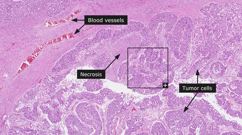
Ovarian cancer
Female, 78 years, poorly differentiated endometroid adenocarcinoma 
Ovarian cancer
Epithelial carcinoma of the ovary is one of the most common gynecologic cancers and the fifth most frequent cause of cancer mortality in women. 50% of all ovarian cancers are diagnosed in women older than 65 years of age. Approximately 5 to 10% of ovarian cancers are familial.
Ovarian cancer is typically denoted as a silent cancer since symptoms occur late in the course of the disease. A majority of ovarian epithelial cancers are detected during physical examination of a pelvic or abdominal mass. By the time of discovery, approximately 70% of the tumors have spread beyond the ovary and are in such cases rarely curable by surgical resection or surgery combined with postoperative chemotherapy and/or radiation therapy. The dismal prognosis has stimulated research efforts for early detection of ovarian cancer.
Ovarian epithelial cancer is bilateral (involving both ovaries) in one-third to one-half of the cases. The FIGO (International Federation of Gynaecology and Obstetrics) staging system recognizes four stages for ovarian cancer. Stage I depicts cancer limited to one or both ovaries. Stage II denotes pelvic extension from ovarian cancers. Stage III cancers show extrapelvic disease and lymph node involvement, and Stage IV tumors show distant metastases. Patients with Stage I tumors have a 5-year survival of 80%, while the five-year survival of Stage IV patients is merely 8%.
Ovarian epithelial cancers are classified into serous, mucinous, endometrioid, clear cell, transitional cell, squamous cell, mixed epithelial and undifferentiated categories depending on histomorphologic features. The most common forms include sero-papillary, mucinous and endometrioid subtypes. Several histologic grading systems have been proposed with the WHO system being widely employed. Grade 1 (well-differentiated) endometrial cancers show less than 5% of solid tumor growth pattern (without lumen formation) and uniform oval nuclei with evenly dispersed chromatin. In Grade 2 cancers, between 6-50% of the tumor is composed of solid masses and nuclei display intermediate features compared to Grade 1 and Grade 3 cancers. In Grade 3 (poorly differentiated) cancers more than 50% of the tumor is composed of solid tumor cell masses and tumor cell nuclei show coarse chromatin and prominent nucleoli.
Normal tissue: Ovary
|