Proximity ligation assay
Proximity ligation assay (PLA) was developed and first described by Fredriksson and colleagues in 2002 (Fredriksson et al., 2002) and became commercialized by Olink Bioscience (http://www.olink.com/). By now it has become an established and generally applicable immunohistochemical tool for advanced and precise protein analysis (Fredriksson et al., 2002; Leuchowius, Weibrecht, & Söderberg, 2011). This innovative method enables exceptional specificity and sensitivity of protein detection and quantification for immunocytochemistry (ICC) and immunohistochemistry (IHC) applications in unmodified cells. In situ PLA allows detecting and counting not only single endogenous proteins but also protein-protein interactions and protein modifications. Selectivity in PLA assays is enhanced by exploiting both antibody and DNA properties; a double recognition by two oligonucleotide-conjugated antibodies provides high selectivity of protein detection, as binding of single proximity probe is not enough to generate signal needed for detection. Moreover the use of DNA and DNA modifying techniques allows detecting and visualizing single protein molecules witch high spatial accuracy and sensitivity.
Technology
The PLA technique utilizes one pair of oligonucleotide labeled antibodies binding in close proximity (30-40 nm apart) to different epitopes of the same protein or two proteins in a complex. The assay is used for localized detection, visualization, and quantification of a single protein or protein-protein interactions in adherent cell lines, cytospin preparations or tissues, including frozen or paraffin-embedded patient samples. The cells or tissue need to be fixed with a fixative appropriate for antibodies used in the PLA protocol and if necessary antigen retrieval and antibody-specific blocking must be performed.
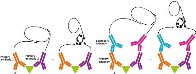
There are two different approaches; one method uses direct primary antibody conjugation and the other uses detection with secondary conjugates, (Figure 1). In the direct PLA method, two primary antibodies that are conjugated to short DNA oligonucleotides, bind a target molecule or interacting molecules. Two additional DNA strands, called connector oligonucleotides, are then introduced. The two DNA strands on the antibodies are ligated to the two additionally introduced connector oligonucleotides, leading to the formation of a circular, single stranded DNA molecule. In the created DNA circle, one of the antibody conjugated DNA probes serves as a primer for the rolling circle amplification (RCA). As a result, when adding a DNA polymerase, a long DNA product is formed and it remains covalently attached to one of the PLA probes. After finalizing the RCA, the concatameric repetitions of the same sequence enable hybridization of multiple detection oligonucleotides that can be visualized under a microscope and quantified (Söderberg et al., 2006). The principle is the same for the indirect form of PLA; however, in this method the unmodified primary antibodies are detected with secondary antibodies that are equipped with DNA strands.
Figure 1. Direct (A) and indirect (B) PLA techniques. The direct method uses antibody pairs with primary conjugation and the indirect method use secondary antibody conjugates.
Specific examples
Applying PLA techniques for proteome screening significantly increases the specificity of in situ protein detection with single-molecule resolution using conventional microscopes and may provide the scientific community with powerful tools for clinical research, protein biomarker validation and routine diagnostics. The improved specificity of protein detection with PLA extends the opportunities for high-performance and large-scale quantitative validation of antibodies, and is used for in situ analysis of antibody functionalization and large scale assays for specific protein expression.
PLA technologies have been used to address various biomedical problems and have shown potential to resolve difficulties during the validation of potential biomarkers for clinical diagnostic needs (Gajadhar, Bogdanovic, Muñoz, & Guha, 2012; Spears et al., 2012; Zieba et al., 2012). PLA has been applied to investigate the role of the epidermal growth factor receptor (EGFR) dimerization and activation during tumor progression and development of drug resistance, and for a more accurate selection of patients for the EGFR-targeted treatment in glioblastoma multiforme (Gajadhar et al., 2012). Another example is the study on the role of human epidermal growth factor receptors (HERs) in early breast cancer. Elevated levels of HER2-HER2 and HER2-HER3 complexes detected by PLA were significantly associated with a decrease in overall survival and reduced recurrent-free survival of breast cancer patients (Spears et al., 2012). The in situ PLA was furthermore applied to detect several proteins, protein-protein interactions, and post-translational modifications in parallel to study simultaneous effects of targeted therapies on cell signaling (Leuchowius et al., 2013; Pinto et al., 2012).
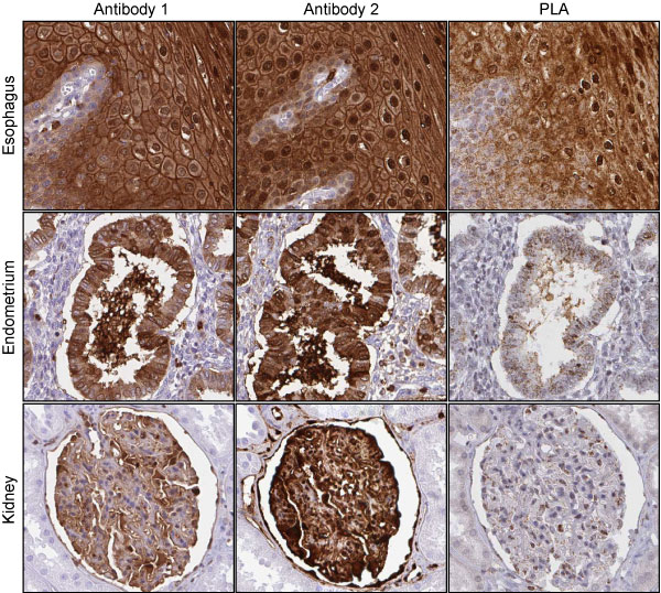
The PLA is employed by Human Protein Atlas (HPA) as an alternative to ICC and IHC, and as a general mean to improve specificity of protein detection on formalin fixed, paraffin embedded cells and tissues. The HPA has developed a robust protocol for the use of PLA; by using mouse monoclonal antibodies in combination with rabbit HPA polyclonal antibodies, the commercially available secondary anti-mouse and anti-rabbit binders with attached oligonucleotides were applied to reveal sites of co-localization of primary antibodies. A new method was optimized for direct conjugation of non-purified, low-concentration antibodies with the PLA oligonucleotide arms for use in the PLA protocols with antibodies derived from the same species. Several pairs of antibodies were applied towards cancer-related protein targets on tissue microarrays (TMAs) consisting of a range of normal and cancer tissues, and also on cell arrays. In addition, the PLA results were validated and confirmed using mRNA sequencing data for the same tissues and cells. Through application of PLA, the specificity of protein detection in comparison to conventional IHC staining was significantly increased, as shown in Figure 2. The established PLA protocols are applicable for studies with HPA reagents on large scale investigations of medical relevance and for biomarker screening.
Figure 2. Expression patterns of Annexin A1 (ANXA1). Antibody 1 (HPA011272) and antibody 2 (CPTC-ANXA1-3) have similar staining patterns in esophagus, endometrium, and kidney. However, when using both antibodies in a PLA reaction only the esophagus exhibits similar detection signals. This correlates well with the detected mRNA level, as the esophagus shows a high mRNA expression of Annexin A1 (TPM 7549), while endometrium (TPM 176) and kidney (TPM 49) have low mRNA expression for this gene.
References and Links
- Fredriksson, S., Gullberg, M., Jarvius, J., Olsson, C., Pietras, K., Gústafsdóttir, S. M., ... Landegren, U. (2002). Protein detection using proximity-dependent DNA ligation assays. Nature Biotechnology, 20(5), 473–7.
DOI:10.1038/nbt0502-473. PubMed: 11981560
- Gajadhar, A. S., Bogdanovic, E., Muñoz, D. M., & Guha, A. (2012). In situ analysis of mutant EGFRs prevalent in glioblastoma multiforme reveals aberrant dimerization, activation, and differential response to anti-EGFR targeted therapy. Molecular Cancer Research: MCR, 10(3), 428–40.
DOI:10.1158/1541-7786.MCR-11-0531. PubMed: 22232519
- Leuchowius, K.-J., Clausson, C.-M., Grannas, K., Erbilgin, Y., Botling, J., Zieba, A., .. Söderberg, O. (2013). Parallel visualization of multiple protein complexes in individual cells in tumor tissue. Molecular & Cellular Proteomics: MCP, 12(6), 1563–71.
DOI:10.1074/mcp.O112.023374. PubMed: 23436906
- Leuchowius, K.-J., Weibrecht, I., & Söderberg, O. (2011). In situ proximity ligation assay for microscopy and flow cytometry. Current Protocols in Cytometry / Editorial Board, J. Paul Robinson, Managing Editor ... [et Al.], Chapter 9, Unit 9.36.
DOI:10.1002/0471142956.cy0936s56. PubMed: 21455970
- Pinto, R., Carvalho, A. S., Conze, T., Magalhães, A., Picco, G., Burchell, J. M., ... David, L. (2012). Identification of new cancer biomarkers based on aberrant mucin glycoforms by in situ proximity ligation. Journal of Cellular and Molecular Medicine, 16(7), 1474–84.
DOI:10.1111/j.1582-4934.2011.01436.x. PubMed: 21883895
- Spears, M., Taylor, K. J., Munro, A. F., Cunningham, C. A., Mallon, E. A., Twelves, C. J., ... Bartlett, J. M. S. (2012). In situ detection of HER2:HER2 and HER2:HER3 protein-protein interactions demonstrates prognostic significance in early breast cancer. Breast Cancer Research and Treatment, 132(2), 463–70.
DOI:10.1007/s10549-011-1606-z. PubMed: 21638049
- Söderberg, O., Gullberg, M., Jarvius, M., Ridderstråle, K., Leuchowius, K.-J., Jarvius, J., ... Landegren, U. (2006). Direct observation of individual endogenous protein complexes in situ by proximity ligation. Nature Methods, 3(12), 995–1000.
DOI:10.1038/nmeth947. PubMed: 17072308
- Zieba, A., Pardali, K., Söderberg, O., Lindbom, L., Nyström, E., Moustakas, A., ... Landegren, U. (2012). Intercellular variation in signaling through the TGF-β pathway and its relation to cell density and cell cycle phase. Molecular & Cellular Proteomics: MCP, 11(7), M111.013482.
DOI:10.1074/mcp.M111.013482. PubMed: 22442258
- A description of the PLA technology:
http://www.olink.com/products-services/duolink/situ-pla-technology
- A video describing the PLA assay:
http://www.youtube.com/watch?v=KbpUU7jQTF8
- Wikipedia - relevant links:
http://en.wikipedia.org/wiki/Proximity_ligation_assay
- Antibodypedia – An open-access database of publicly available antibodies and their usefulness in various applications:
http://www.antibodypedia.com
|
 The Human Protein Atlas project is funded


 MENU
MENU